|

Figure Drawing EbooksThe Art Student's Guide to External Anatomy |
Learn How to Draw Perspective
Purchase the ebook by clicking the "Buy Now" button at the bottom of the page.
After completing the purchase you will be directed to a web page which will give you a link to the download site.
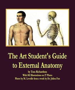
The Art Student's Guide to External Anatomy
  See a paperback edition of The Art Student's Guide to External Anatomy Or preview a copy at Google Books |
Anatomy for Artists Painters and Sculptors

|
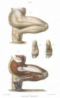
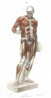
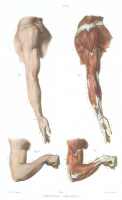
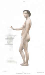
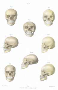
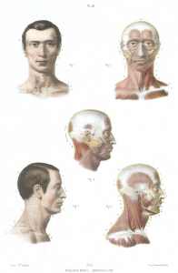
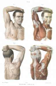
Anatomical Drawings for Art Students, Painter and Sculptors in full color, with additional ink drawings of bones and muscles. This is a reprint of a nineteenth century Atlas of lithographs by M. Leveille which accompanied the other volume of descriptive text by Dr. Julien Fau. |
|
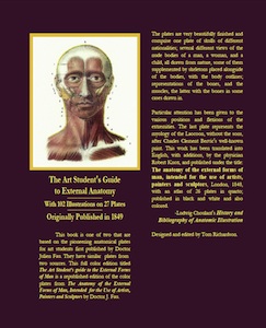
About this ebook This is a republished edition of The Anatomy of the External Forms of Man, Intended for the Use of Artists, Painters and Sculptors by Doctor J. Fau, Lithographed by M. Leveille', pupil of M. Jacob, New edition with additional plates by William Norris. I located another edition with a publication date of 1849. The following information about Dr. Fau, the editor of the book is from Ludwig Choulant's History and Bibliography of Anatomic Illustration, published in 1920. JULIEN FAU Julien Fau, doctor of medicine in Paris, edited two different anatomic works for artists: Anatomie des formes exterieures du corps humain, a l'usage des peintres et des sculpteurs. Avec un Atlas de 24 planches dessinées d'apres nature et lithographiees par M. Leveille, eleve de M. Jacob. Paris: Mequignon- Marvis fils, 1845 (16 and 214 pp.), 8° and fol. (24 lithographic plates), black and white and also colored.
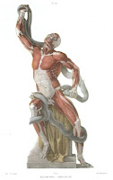
Particular attention has been given to the various positions and flexions of the extremities. The last plate represents the myology of the Laocoon, without the sons, after Charles Clement Bervic's well-known print. This work has been translated into English, with additions, by the physician Robert Knox, and published under the title: The anatomy of the external forms of man, intended for the use of artists, painters and sculptors, London, 1848, 8°, with an atlas of 26 plates in quarto; published in black and white and also colored.
|
The second, smaller, and less expensive work by Fau is: Anatomie artistique elementaire. Dessins d'apres nature par J. B. Leveille, gravures sur acier. Paris: Mequignon-Marvis, 1850, 8°, with 17 steel engravings in 8°, three of which are in small folio.In this work the representation of the shapes of skulls, of the nude bodies, and of the Laocoon are missing, but representations of three beautiful skeletons, with the contours drawn around them, have been added. The remainder deals with osteology and myology, although less exhaustively than in the previous work. There were as many as ten editions of the book published in French, English, Dutch and German. The Anatomie artistique elementaire (Petit Fau) has shortened descriptions and only 17 steel engraved plates. I have come across an English version of it with additional illustrations and notes on sculpting, muscular anatomy and human proportion. I am working on an ebook version of this. Below are 3 photos of the French Edition from 1865. This was a self published book by Jonathon Scott Hartley which contains the same steel black ink engravings as Anatomie artistique elementaire . Hartley's book combines the plates with extensive commentary on anatomy and a chapter on proportion and the art of modeling (sculpting in clay). Another English edition with plates was Elementary Artistic Anatomy of the Human Body, Translated and edited by Charles Carter Blake was published in 1881. There is a reference to Carter Blake in Charles Darwin's letters, Blake was an advocate of the discredited study of cranioscopy, which tried to make sense out of the races by a study of skulls. There was a schism in Victorian science with some arguing that cranioscopy proved distinct races, and ethnologists arguing for the unity of the races.
F. von Hebra informational website with images taken from Anton Elfinger's lithographs for Hebra's book. Additional notes below and more notes at my blog: Figure Drawing: Notes, Illustrations and Books about Figure Drawing and the Proportions of the Human Form. |
DOWNLOAD THE E-BOOK The Art Student's Guide to External Anatomy
82 pages with 27 plates and 23 additional illustrations
plus an addendum of 12 ink drawings of bone and muscle structure.
- $14.95
|
DOWNLOAD THE E-BOOK Buy now with Paypal. If you are new to PayPal you will be directed to a PayPal sign up page or you will be allowed to pay directly by credit card. |
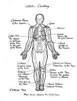
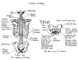
You will need Adobe Acrobat Reader (c) to view the PDF file. If you do not have a copy of Adobe Acrobat Reader you may download a free copy of the latest version here: Acrobat Reader Download Site If you experience any trouble downloading the e-book please click on this link: Detailed Download Instructions If you are still experiencing trouble email me at:lifedraw2008@gmail.com and I will contact you to help with the download or email you the file. Included in the e-book is an additional section of anatomical pen and ink drawings illustrating the bones and muscular structure of the human body, examples are shown below. Because of the cost of full color printing I left these black and white drawings out of the printed Amazon.com edition. If you purchase the printed book and wish to have these drawings as a pdf file, email me with a proof of purchase, and I will send you a link. |
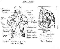
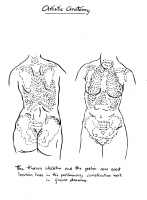
|
This is a republished edition of The Anatomy of the External Forms of Man, Intended for the Use of Artists, Painters and Sculptors by Doctor J. Fau, Lithographed by M. Leveillé, pupil of M. Jacob, New edition with additional plates by William Norris. I located another edition with a publication date of 1849. The same author had a young artist, Eugene Caudron, a pupil of David d'Angers, make an anatomic plaster statuette, 70 centimeters high, for the use of artists (nouvel ecorche) which is sold, white or colored, with a description and four pages of pictorial representations (the four views of the statuette), for 15 and 30 francs respectively (the description alone costs 3 francs). Prior to this, Johann Martin Fischer's plaster.
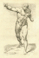
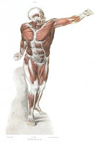
Preceeding Dr. Fau's book and perhaps influencing it was another by P.N. Gerdy similarly titled Anatomie des Formes Exterieures which included 3 schematic diagrams of the human form. Another book with art designed by M. Leveille and engraved under his supervision was Heath's Operative Surgery. A Course of Operative Surgery, consisting of a Series of Colored Plates, each plate containing Several Figures, Drawn from Nature by the Celebrated Anatomical Artist, M. Leveille, of Paris, Engraved on Steel under his immediate superintendence, with Descriptive Text of Each Operation, and numerous Wood Engravings. By Christopher Heath, F.r.c.s., Surgeon to University College Hospital, and Holme Professor of Clinical Surgery in University College, London. One Large Quarto Volume. Second Edition, Revised and Enlarged. Sold by Subscription. Full information upon application. Cloth, $12.00. P. BLAKISTON, SON & CO., 1012 Walnut St., Philadelphia. Skin in Water-Colours: Unpublished Aquarelles from Hebra's Department in Vienna 1841-1843The last three plates in the book are dissections of ancient statuary, The Laocoon, The Gladiator and Discobolus. Link to Washington School of Medicine - Monuments of Medicine page - rare books from the Bernard Becker Medical Library, which has a note on the French edition and a picture of the Laocoon Plate, compare it to the original sculpture. The Laocoon plate was original to the French edition, the Gladiator and Discobolus are original to theEnglish edition. Anatomie Des Formes Exterieures Du Corps Humain A link to an exhibition of Jean Baptiste François Leveillé's works and another one and a link to the home page of the exhibition Anatomy Acts. Google Books result for Traite Practique D'Anatomie, with plates by Leveillé. According to Boris Röhrl in his History and Bibliography of Artistic Anatomy, Julien Fau's textbook along with Duval's Precis d' Anatomie, were the most successful publications of their period. Fau's invention was to supplement the textbook with the large atlas of prints. Röhrl also notes that for the reader not interested in the extensive description of the prints found in the textbook a pocket book, Anatomie artistique elementaire, was created. French at students called the small volume Petit Fau, and the large textbook Grand Fau. Röhrl argues that while Fau was influential in anatomical studies, his main concern was with the faithful rendering to produce art. "The only question posed is why a picture executed with the greatest scientific exactitude should not be an effective work of art at the same time.Thoughts based in the metaphysical or spiritual sphere were not considered by Fau, but he does recognize that the plain reproduction of nature did not produce artistic value in a certain work to the same degree. His main argument lies in a continuation of the tradition of the Renaissance." You can search for a copy of Röhrl's book at Abebooks.com. Use the search terms "Boris Röhrl," or "History and Bibliography of Artistic Anatomy." An English translation of the large textbook (Grand Fau) without the plates is at Google Books The Anatomy of the External Forms of Man, ed. with additions by R. Knox. A reprint of the atlas of anatomical prints is what makes up my book, Anatomy for Art Students, Painters and Sculptors. Links: Bibliothèque artistique de la Ville de Bruxelles - Art Library of the City of Brussels - Image from Anatomie des formes exterieures du corps humain.Artist versus Anatomist, Models against Dissection: Paul Zeiller of Munich and the Revolution of 1848 - Article by Nick Hopwood, MSc, PhD - mentions Anton Elfinger's employment.
These arrangements were informal, but then "anatomical modeller" was not a regular occupation. Medical models were made by artists, preparators and others. Most German universities had a drawing teacher, but professors' attempts to create posts for scientific artists tended to founder on the combination of demanding job description and low academic and artistic status. Models were more of a luxury than drawings and the skill was less widely distributed. Some state collections already employed staff to prepare specimens for display‹Munich's zoological preparator had a doctorate‹but it was rare for a medical modeller to gain a dedicated position, such as Joseph Towne enjoyed at Guy's. In Vienna Dr Anton Elfinger was from 1849 hired by the medical faculty to produce illustrations and models, but the money soon ran out. A little later at the University of Freiburg in Baden Dr Adolf Ziegler made embryological waxes as a zootomical Assistent, a position that for others was a stepping-stone to a chair, but from which he resigned in 1868 to build up a private studio. Links: Skin in Water-Colours: Unpublished Aquarelles from Hebra's Department in Vienna 1841-1843 Cajetan das Leben des Wiener Mediziners und Karkaturisten Dr. Anton Elfinger The Artist's Guide to Human Anatomy Images from an 1880 edition are posted at this blog: Autumn Cottage Diarist Days in the life of an English country woman - Anatomical Atlases and Multi-Layered Men |
This new edition is copyright 2010, the original images in it are believed to be in the public domain based on their age and publishing date. If you have information to the contrary please email me: lifedraw2008@gmail.com
Also available in a previous edition, please visit Lulu.com |


